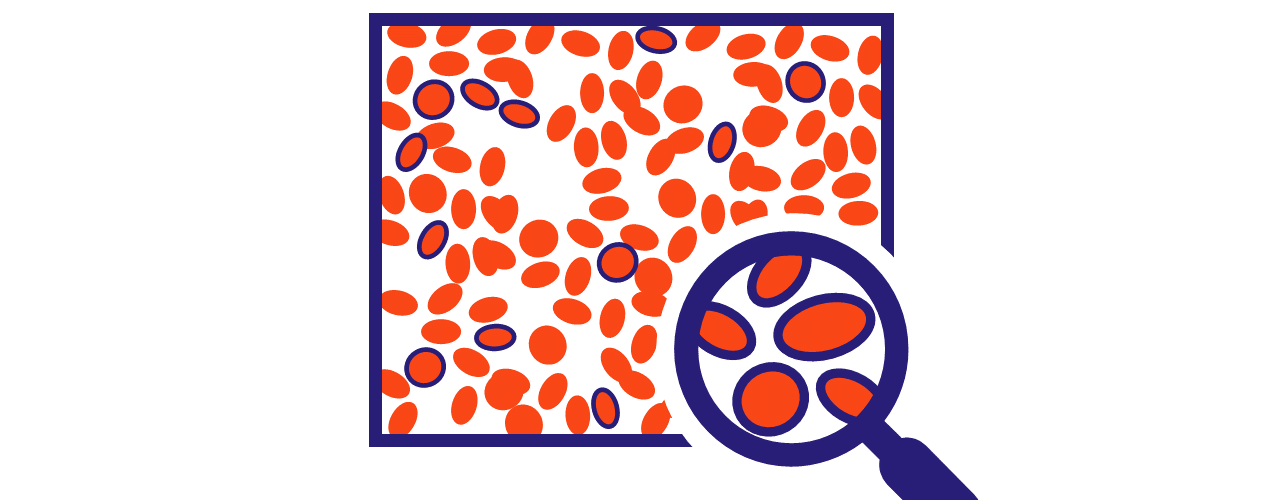
The image analysis division develops and maintains pipelines and interfaces to visualize, process, and analyze a wide range of biomedical imaging data, providing scalable and reproducible workflows. This includes utilizing image processing and advanced deep and machine learning techniques for tasks such as segmentation, feature extraction, and classification. Additionally, the division continuously adapts to emerging technologies like spatial transcriptomics (e.g., 10x Visium and Vizgen MERSCOPE). For more information please contact us at dac_image@icm-institute.org.
Image patching & stitching
Some images can be too large to process on a whole-image level, and may need to be broken down into smaller patches. Once these patches are processed, they might be required to be “stitched” back together. Most existing software is slow and computationally expensive. We provide solutions at a fraction of the time.
Automated quality assessment
Automated quality assement provides a fast and objective way to detect images or regions of poor quality. Visual markers can be superimposed on regions that have a high likelyhood of poor quality across several well-established statistics.
Filtering & Enhancement
Images can be enhanced (e.g., based on quality assessment), e.g. by removal of blurry regions, removal of salt and pepper or granulated noise, correction of illumination, or resolving of contrast issues. The solutions range from traditional image processing techniques to advanced methods, such as deep learning models.
Segmentation
The goal of segmentation analysis is to precisely identify specific regions of interest in microscopy or neuroimaging images. These can go from single nuclei, to cell membranes (oligodendrocytes, astrocytes or microglia), tumors, all the way up to whole brain regions in MRI. Many approaches can be used, depending on the question, from using traditional image processing to machine learning models. The results of segmentation are binarized or multi-layered masks, visualization on the original image for validation, and tabular data summarizing segmentation statistics such as count and locations.
Feature extraction & statistics
The goal of feature extraction is to quantify descriptive features that allow correlations with conditions or outcomes. These features can include metrics such as intensity, size, shape, radiomics, and complex shape-related characterizations that can quantify the degree of branching. To further exploit the potential for explanatory information, a broad analysis approach can be performed, applying a large battery of feature extractions that are then ranked according to their explanatory value for further statistical applications.
Image registration & reconstruction
We can align time-series or stacked 2D images that represent a 3D structure. Precise alignment is crucial, especially for tracking disease progression over time, such as monitoring tumor changes. For 3D reconstruction, accurate image registration ensures that the final model matches the original structure. In addition to aligning images, we also provide quality metrics, such as the similarity measurement index, to assess the accuracy of the alignment.
Visualization
We provide solutions for visualizing and interacting with 2D and 3D images through custom-made modules, delivered as scripts, macros or modules in existing software. Available tools include FIJI/ImageJ, QuPath, OMERO, and Vizgen postprocessing.
Automation
Manual processing of images can be error prone, time-consuming and difficult to reproduce. We provide expertise in automating most image processing steps, including (meta)data management, in collaboration with our Data Governance & Open Science team, and the DSI

